Imaging Services
To help physicians diagnose illness in its earliest and most treatable stage, Randolph Health has an array of state-of-the-art imaging services. These services are used to detect major and minor conditions, from a blood clot in the brain to a broken bone or skull fracture. Qualified and skilled physicians and technologists use the most up-to-date technology to provide you with the best diagnosis for treatment.
Our board-certified radiologists, registered radiologic technologists and staff are committed to providing the best care available in a warm and compassionate environment. Our services include:
Computed Tomography (CT)
CT imaging is particularly useful because it can show several types of tissue—lung, bone, soft tissue and blood vessels—with great clarity. Using specialized equipment and expertise to create and interpret CT scans of the body, radiologists can more easily diagnose problems such as cancers, cardiovascular disease, infectious disease, trauma and musculoskeletal disorders.
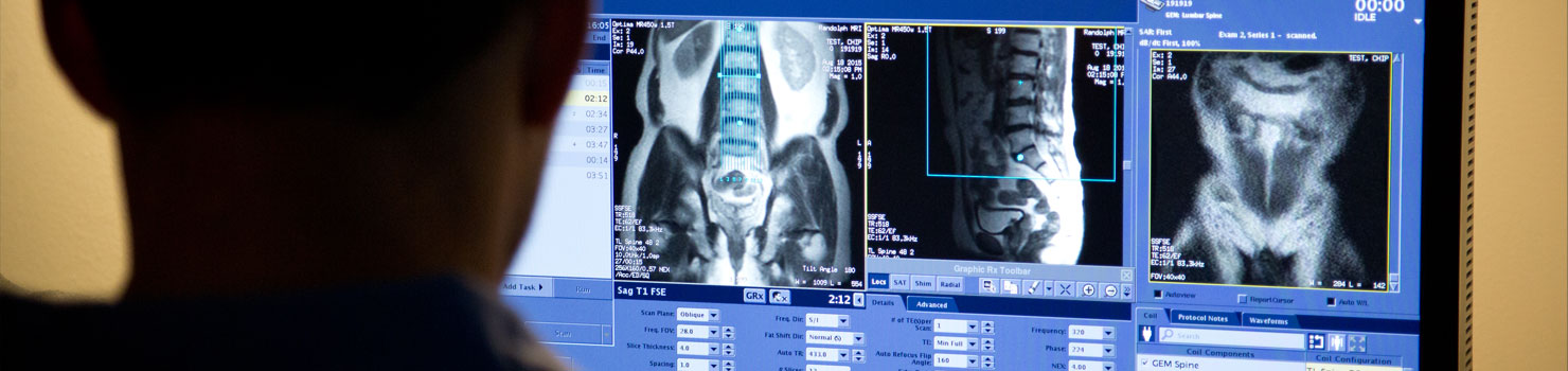
Magnetic Resonance Imaging (MRI)
Because MRI can give such clear pictures of soft-tissue structures near and around bones, it is the most sensitive exam for back, neck and joint problems. MRI is widely used to diagnose sports-related injuries, especially those affecting the knee, shoulder, hip, elbow and wrist.
Breast Services
The Breast Center at Randolph Health offers total care for your overall breast health. Using innovative medical technologies and procedures designed specifically for women, such as digital mammography and stereotactic breast biopsy, the Breast Center at Randolph Health is your destination for all your breast health needs. Board-certified radiologists, state-of-the-art digital mammography equipment, high-resolution ultrasound and image-guided biopsy services are just a few of the amenities offered at the Breast Center. Our comfortable facilities offer private dressing rooms and convenient scheduling.
Our comprehensive Breast Center allows women to get results fast and conveniently schedule any additional testing needed, such as diagnostic mammography, ultrasound or biopsies.
Nuclear Medicine
Nuclear medicine procedures use radioactive materials to perform body function studies and organ imaging, analyze biologic specimens, and treat disease.
Radiography (x-ray)
Radiography, known to most people as x-ray, is most commonly used to assist the physician in identifying and treating fractures. X-ray images of the skull, spine, joints and extremities are performed every day in hospital emergency rooms, sports medicine centers, orthopedic clinics and physician offices. Images of the injury can show even very fine hairline fractures or chips, while images produced after treatment ensure that a fracture has been properly aligned and stabilized for healing. Bone x-rays are an essential tool in orthopedic surgery, such as spinal repair, joint replacements or fracture reductions.
Ultrasound
Ultrasound, or sonography, involves the sending of sound waves through the body. Those sound waves are reflected off the internal organs. The reflections are then received by special instruments that subsequently create an image of anatomic parts. No radiation is involved in ultrasound imaging.